

We have all been there, neglected a niggle in our foot and told ourselves that it would probably go away. Weeks have passed, even months, and it’s only getting worse. Then finally, when we are hobbling around barely able to walk, we decide to seek help…
Well, at Achilles Foot Clinic, we are here to help you.
Seeing Clearly vs. Seeing in Motion: MRI and Ultrasound Compared for Heel Pain
MRI vs. Ultrasound: Decoding the Diagnostic Dance for Heel Pain
Heel pain. It can be a relentless foe, impacting your daily activities and leaving you wondering – what's causing this discomfort? Here at Achilles Foot Clinic, we understand the importance of an accurate diagnosis for effective treatment. Today, we'll delve into the world of diagnostic imaging, specifically comparing two common techniques used for heel pain: MRI and Ultrasound.
MRI: A Peek Inside
Magnetic Resonance Imaging (MRI) is a powerful imaging technique. It utilises strong magnetic fields and radio waves to create detailed cross-sectional images of your foot and ankle. This allows podiatrists to visualise:
Bone abnormalities: MRI excels at revealing fractures, stress fractures, and bone marrow edema (inflammation) within the heel bone.
Ligament and tendon tears: Tears in the plantar fascia, Achilles tendon, or other ligaments can be clearly identified with MRI.
Joint problems: MRI can detect abnormalities in the ankle joint, such as arthritis or cartilage damage.
Advantages of MRI:
Unparalleled detail: MRI offers the most comprehensive view of the soft tissues and bones within the foot and ankle.
Non-invasive: The procedure is painless and doesn't involve radiation exposure.
Limitations of MRI:
Cost: MRI scans are typically more expensive than ultrasounds.
Accessibility: MRI machines are not as readily available as ultrasound equipment.
Claustrophobia: The enclosed nature of the MRI scanner can be an issue for some patients.
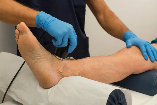
Ultrasound: A Real-Time Look
Ultrasound imaging uses high-frequency sound waves to create real-time images of your foot and ankle. This technique is particularly useful for:
Dynamic assessment: Ultrasound allows visualisation of structures in motion, such as tendons and ligaments, during movement. This can be helpful in identifying tears or inflammation.
Targeted guidance: Ultrasound can be used to guide needle placement for injections like cortisone shots.
Advantages of Ultrasound:
Cost-effective: Ultrasound is generally less expensive than MRI.
Portable: Ultrasound machines are often portable, allowing for more convenient examinations.
Real-time visualisation: The ability to see structures in motion provides valuable insights.
Limitations of Ultrasound:
Limited view: Ultrasound doesn't penetrate as deeply as MRI, making it less effective for visualising bone details.
Operator dependence: The quality of ultrasound images relies heavily on the skill of the technician performing the scan.
Choosing the Right Diagnostic Tool
The choice between MRI and an ultrasound depends on the specific suspected cause of your heel pain. Here's a simplified breakdown:
For detailed visualisation of bones, ligaments, tendons, and joint structures: MRI is the preferred choice.
For evaluating soft tissue movement and real-time guidance for injections: Ultrasound is a good option.
Ultimately, your podiatrist will determine the most appropriate imaging technique based on your individual case and medical history.
Don't let heel pain hold you back! At Achilles Foot Clinic, our experienced podiatrists utilise advanced diagnostic tools and their expertise to get you on the path to a pain-free future. Contact us today to schedule an appointment!
Ask Lorcan And His Team
Fill in the form to request a Call From Our Team
Fill in the form to request a Call From Our Team
One of our team will call you for FREE and answer any questions or concerns you may have about Bunions.
One of our team will call you for FREE and answer any questions or concerns you may have about your uncomfortable Bunions.

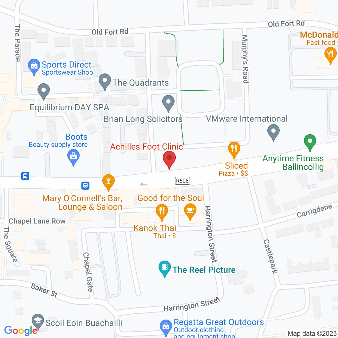
Where To Find Achilles Clinic
Ready to take the next step?
We are conveniently located in the western side of Cork City in Ballincollig.
Main Street, Ballincollig
Open 8am – 6pm (Mon-Fri)
Free Parking on our doorstep
We are on the 220 bus route


Legal
How to get in touch
© Copyright 2022. Achilles Foot Clinic. All rights reserved.

